01 / THE PATIENT ARRIVES
Airway secured, bilateral mandibular destruction still to solve.
The patient presented after a facial gunshot wound. Emergency tracheostomy had already secured the airway. Examination documented lacerated and contused facial wounds, a projectile entry wound and communication with the oral cavity.
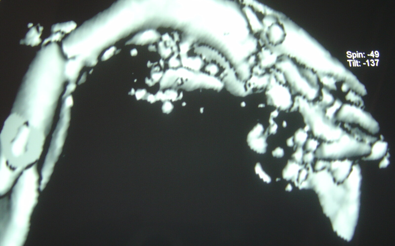
The skeletal injuries included multiple bilateral mandibular fractures with simple, compound and comminuted components, a mandibular alveolar fracture, dental avulsion and loss of bone on the left side.
02 / THE WORKING DIAGNOSIS
One mandible, two different mechanical and biological environments.
The right side required dependable stabilization to re-establish mandibular continuity. The left side combined comminution, bone loss and direct oral exposure, creating a substantially greater contamination risk and a poor environment for extensive hardware placement.
The remaining teeth and occlusal relationship were limited but clinically valuable. They offered a reference for restoring mandibular position and supporting a less invasive strategy on the left.
The case was treated in June 2006. The surviving image series contains assessment, operative and postoperative records from different dates within that month.
03 / POSSIBILITIES AND DECISION
Treat each side according to what it could safely support.
The team considered rigid internal fixation for the right-sided fractures and a more conservative, semi-rigid strategy on the left, using the occlusion and remaining teeth as positional references. The existing tracheostomy facilitated maxillomandibular fixation without competing with the immediate airway plan.
Extensive exposure and rigid hardware placement across the contaminated left defect could have increased infection risk and further disrupted the tissues surrounding the area of bone loss. The chosen plan therefore combined surgical stability on the right with protection of the biologic and muscular envelope on the left.
04 / OPERATIVE RECORD
Occlusion-guided fixation plus selective open reduction.
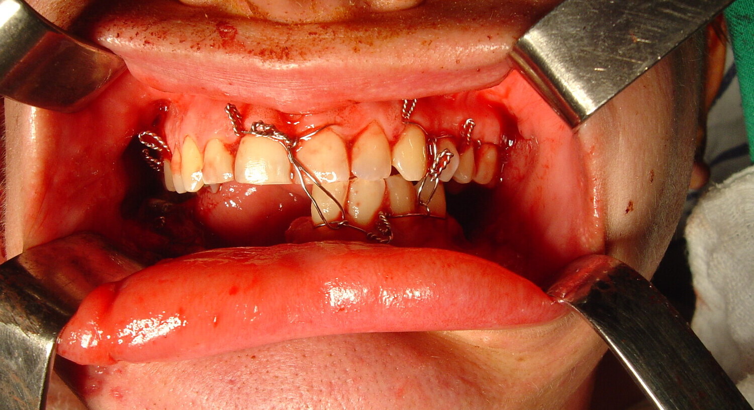
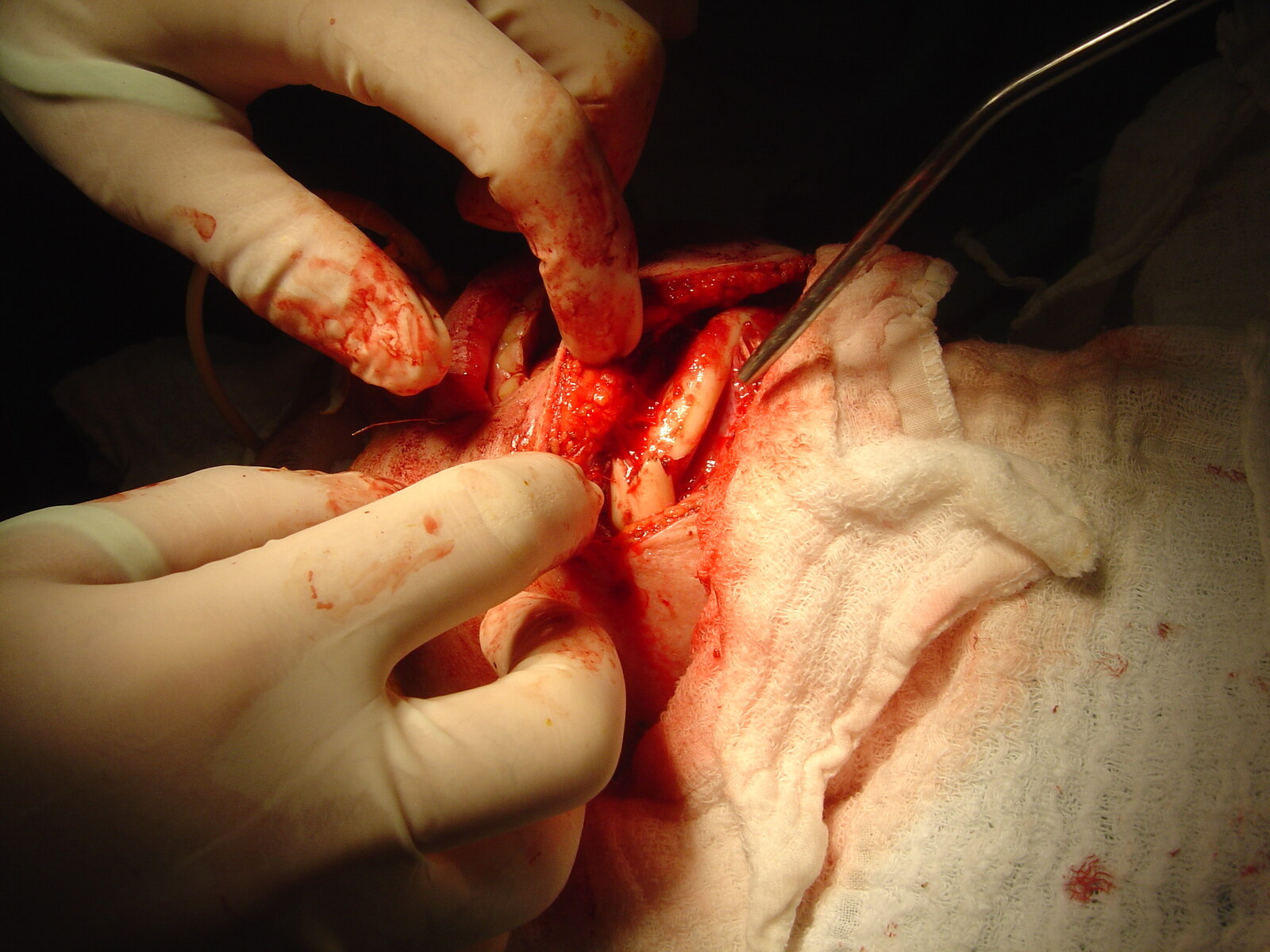
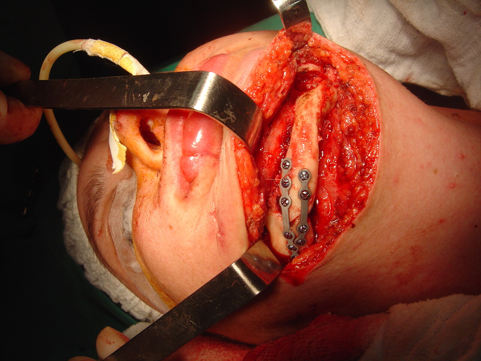
Maxillomandibular fixation used the remaining dentition to guide the occlusion despite the limited number of mandibular teeth. On the right, an extraoral approach exposed the fractured mandibular body and angle. The fragments were reduced and stabilized with rigid plate fixation.
The left side was maintained through the occlusion-guided, less invasive strategy described by the curator. The objective was to preserve the early hematoma, inflammatory response and soft-tissue envelope while avoiding additional contamination and devascularization at the site of bone loss.
05 / RECOVERY AND LONG-TERM OUTCOME
No additional operation and no recorded complication.
The postoperative course required no additional surgery, and no complication was recorded in the submitted history. Early follow-up documented a functional and esthetic occlusion.
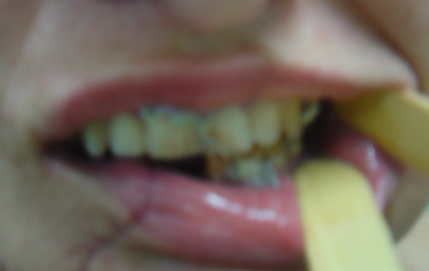
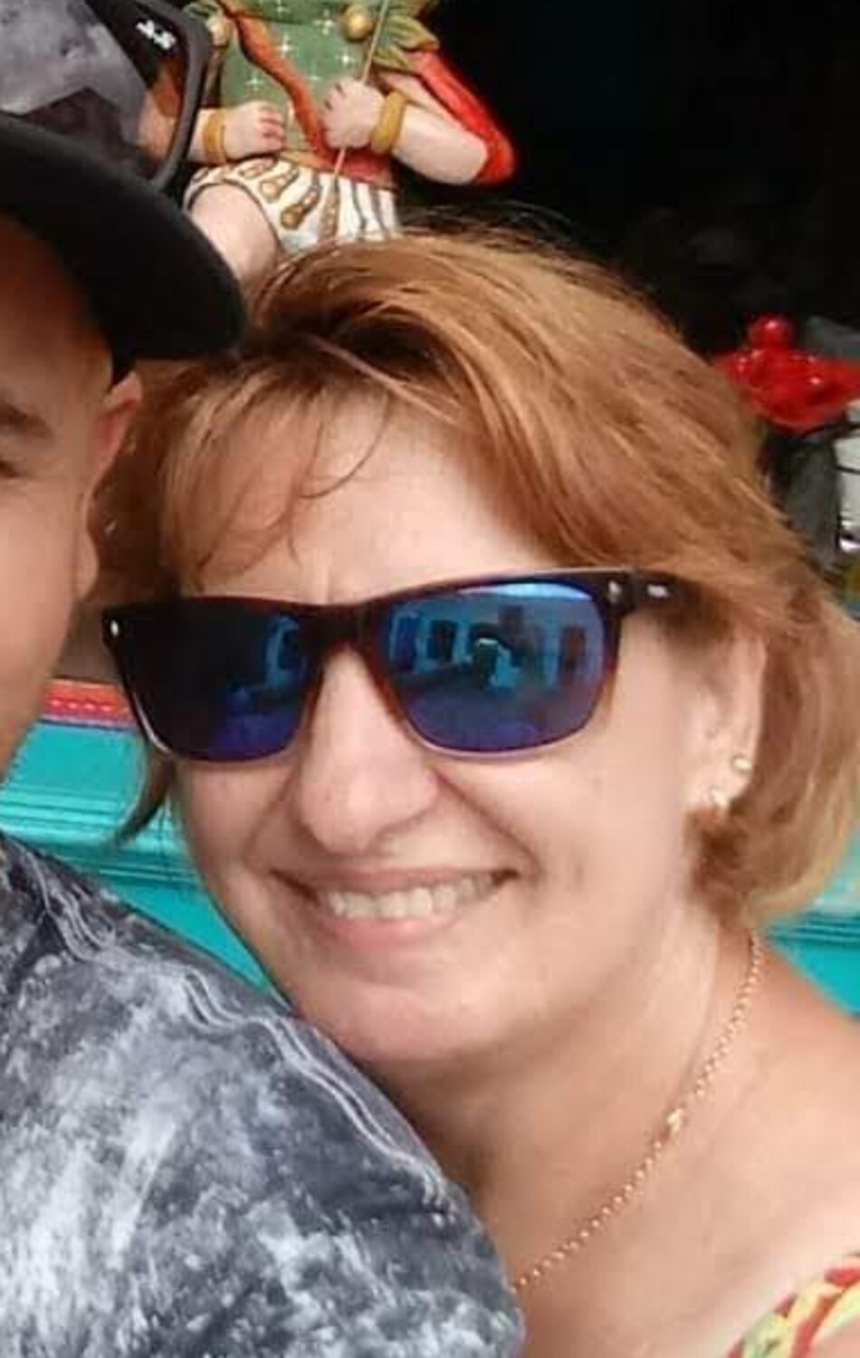
The curator reports that bone formation and remodeling were complete after one year. At 15 years, the patient remained functionally and esthetically rehabilitated and had subsequently received implant and prosthetic rehabilitation.
06 / WHAT THE TEAM LEARNED
Conservative and invasive treatment were complementary, not contradictory.
In the team's interpretation, the early clot and inflammatory response helped maintain the relationship between the injured bone and surrounding musculature on the left. Preserving that environment may also have reduced cicatricial muscle retraction associated with the osseous defect.
The case supports a broader principle from Professor Lasco's school: the most aggressive reconstruction is not automatically the most appropriate treatment for every region of the same injury. Stability, contamination, remaining anatomy, occlusion and biological preservation must be balanced separately.
With contemporary imaging, fixation systems, virtual planning and rehabilitation resources, the tools available today would be better. The central decision could still be similar: rigidly stabilize the side that requires and supports fixation while treating the contaminated comminuted defect according to its biological conditions.